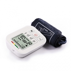
- Descripción
- información opcional
Configuración estándar
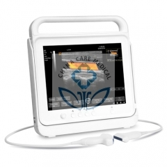
Unidad principal LCD de 1, 15 pulgadas sin sondas;
2, una sonda enchufes;
3, USB, VGA, VIDEO, PRINT, puertos dicom;
4, Pw, thi, tdi, itouch, dopper de color;
5, sistema Windows xp completamente digital
6, 18 meses de garantía Disco duro de 64G
7, sonda convexa
Modo de visualización
Modo B / N: B, 2B, B / M, B / C
Modo Doppler color: CFM, PDI, PW
Dúplex: 2D simultáneo en tiempo real, Doppler
Escala de grises: 256
Pantalla: monitor LCD de 15 pulgadas
Frecuencia del transductor: 2,5-10 MHz
Tecnología digital: enfoque dinámico de recepción (DRF)
Escaneo de frecuencia dinámica (DFS)
Profundidad de escaneo: 300 mm
Procesamiento de imágenes
Preprocesamiento: TGC de 8 segmentos
Preestablecido
Ganancia (blanco y negro, color, Doppler)
Audio
PRF
Gama dinámica
Mejora de la imagen
Potencia Acustica
Post-procesamiento: mapa gris
Reverso blanco / negro
Reversa izquierda / derecha
Arriba / Abajo Reversa
Funciones:
Cine-loop: memoria de bucle de cine de 1000 cuadros
Medios de almacenamiento: capacidad masiva de almacenamiento de imágenes de 64 G
Zoom: Zoom panorámico
Puertos USB: 2
THI: imagen armónica de tejidos
Yo toco
Imágenes claras
Medición y cálculos
Modo B: distancia, circunferencia, área, volumen, ángulo, crecimiento fetal, cruve
Modo M: distancia, tiempo, velocidad, frecuencia cardíaca
Paquete de software: abdomen, ginecología, obstetricia, cardiología, piezas pequeñas, urología Otros
Conectores de transductor: 1 puerto de conector de transductor activo
Puertos periféricos: video, S-video, VGA, 2 puertos USB, puertos Diom
Fuente de alimentación: 100 ~ 240VAC + _10%, 50Hz / 60Hz
Transductores:
Transductor de matriz convexo electrónico
Transductor de matriz transvagional electrónico
Transductor de matriz lineal electrónico
Transductor electrónico microconvexo
| Focusing | Electronic focusing + acoustic lens focusing Transmission focus: single focus, double focus, 3-focus and 4- focus Continuously dynamic focusing mode is used for front end reception |
| B-type display | Image amplification (real-time or freeze status), max. 4-times |
| Image vertical / horizontal overturn | |
| Display depth is continuously adjustable |
|
| M-type display |
Scanning speed: 4- stage |
| Digital beam formation | Image whole range continuous dynamic focusing Image whole range dynamic aperture Image whole range dynamic trace change Image whole range receive delay weight summation Support semi-moment scanning, and support ± 10 ° linear reception deflection angle Multiple beam parallel processing technology |
| Signal processing and Doppler | Provided with dynamic filtering and orthogonal demodulation Provided with total gain adjustment Gain adjustment: 8-segment TGC B-type, C-type, D-type total gain, respectively adjustable B/W image gain and color bloodflow gain, respectively adjustable Doppler stereo output volume adjustment : 0-255 Doppler baseline adjustment : 6-stage Pulse repetition frequency can be respectively adjusted: CFM PWD Provided with D-linear speed adjustment |
| Image processing function | Image realizes dynamic range conversion and logarithm compression Image realizes time filtering Image realizes spatial filtering Image realizes frame correlation Image realizes edge enhancement Image realizes grey scale convertion Provided with selection of B|M-type or M-type scanning speed Provided with high-density and high frame frequency selection Provided with image convex array scanning angle, linear array scanning depth control Provided with image optimized processing: 6-stage Image left/right, up/down overturn Color grey scale bar inversion selection B/W image resolution 1024 × 768 × 8 bit (256 grey scale) Color image resolution: 1024 × 768 8 bit × 8bit × 8 bit color coding |
| Basic measurement Calculation function |
B mode basic measurement: distance, angle, perimeter and area(ellipse method, trace method), volume, stenosis, column diagram, cross-section diagram |
| M-mode basic measurement: heart-rate, time, distance, speed | |
| Doppler measurement: time, heart rate, speed, acceleration |
|
| Obstetrics measurement Calculation function |
Gestational sac(GS), biparietal diameter (BPD), cephalo-rump length(CRL), femur length (FL), Humerus length (HL), Transverse Abdominal Diameter (TAD), backbone length (LV), occipitofrontal diameter (OFD), Abdominal circumference (AC), head circumference (HC), estimation of parietal transparent length, gestational age and expected date of childbirth. |
| Obstetrics report | Measure and calculate amniotic fluid index(AFI) Calculate the rate (BPD/OFD, FL/AC, FL/BPD, HC/AC) Estimate fetus weight Infer the gesattional weeks and expected date of childbirth according to LMP , BBT Fetal Biophysical Score Fetus growth curve |
| Gynaecology measurement Calculation function |
Measurement and calculation of uterus, left ovary, right ovary, left follicle, right follicle etc |
| Andrology measurement Calculation function |
Measurement and calculation of prostate, testicle etc Calculation of prostate peculiar antigen predicted value PPSA, and prostate peculiar antigen density PSAD |
| Urology measurement Calculation function |
Measurement and calculation of left kidney, right kidney, bladder, residual urine volume etc. |
| Peripheral blood vessel measurement Calculation function |
Measurement and calculation of area stenosis rate, tube diameter stenosis rate |
| Small parts measurement Calculation function |
Measurement and calculation of thyroid, breast , enclosed mass etc |
| Cardiac measurement Calculation function |
Cardiac measurement software packet – providing analysis and measurement method for heart rate, speed, left ventricle, aorta, mitral valve, ventricle (right/left) Area stenosis rate percentage (%Area Sten), tube stenosis rate percentage (%Diam Sten) Body surface area (BSA). |
| Memory Function | Probe parameters memory Image memory Cine memory Measurement result memory Report memory |
| Cine function | Auto playback Manual playback Playback speed selection Retrieval playback Forward/reverse playback frame by frame |
| Reports | Obstetrics report Gynaecology report Uology report Andrology report Cardiac report (left ventricle, aorta, mitral valve, ventricule ) Add ultrasonic imgae in report Directory management File management |
| Grey scale |
256 Grey scale |
| Input/Output interface |
Network interface USB interface Video interface Serial communication interface |
| Probes | Probe socket: 1 PCS Probe frequency: 2.5MHz~10.0MHz, 8-stage multi-frequency |
| Probe class |
Large convex array, micro- convex array, high-frequency linear array, transvaginal probe |
| Software upgrading functions |
Available |
| Liquid Crystal Display |
SVGA line-by-line, flashless,15 inch high resolution LCD |
| Power supply |
Built-in switching power supply: input power ≤150VA Power supply adaption range: 220V ± 10%,50Hz ± 1Hz (AC) |
Opcional
Sonda lineal de alta frecuencia de 7.5Mhz
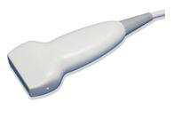
Sonda de matriz microconvexa de 5.0Mhz
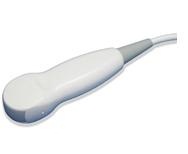
Sonda transvaginal de 7.5 Mhz
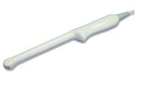
Sonda transrectal de 7.5Mhz
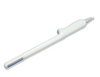
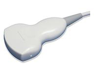
Sonda de matriz convexa de 3,5 Mhz
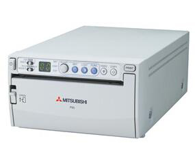
Impresora
Puede comprar cualquier impresora de video en su mercado nativo.
Sugerimos que el modelo sea la impresora de video de la marca Mitsubishi P93C
Shipping Information:
10kg
0.5m,0.3m,0.5m
Piece
No
 USD
USD EUR
EUR GBP
GBP CFA
CFA
